Brain Power
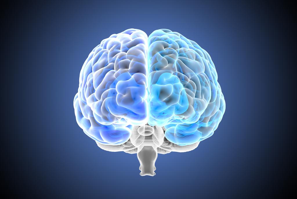
Dr. Yaara Erez decodes brain signals from various imaging techniques and attempts to understand how they relate to functioning and thinking processes. Her breakthrough research made it all the way to the BBC.
In October 2018, British TV channel BBC World aired a segment about an innovative medical procedure: neurosurgery, performed on conscious patients. The patient in the segment, 29-year-old Ben Rush, had a non-aggressive brain tumor that needed to be removed. These types of tumors tend to merge with the healthy tissue around them, so the challenge included removing the tumor without damaging brain functions, to ensure Ben the best possible quality of life and level of functioning. What was special in Ben’s surgery was that during the surgery he also participated in the research, aimed at developing new techniques for this procedure. This is where Dr. Yaara Erez, who specializes in brain mapping and led this research, enters the picture: during the surgery, she asked Ben to perform a series of cognitive tasks such as counting or repeating sequences of letters or numbers, while special electrodes were connected to his brain. “This method, called electrocorticography (ECOG), allows us to locate significant signals via the direct recording of activity from the exposed brain,” explains Dr. Erez. “Our research data showed that there are indices that can tell us which areas of the brain are relevant or related to different cognitive functions.”
Dr. Erez, married +3, has been mapping brain functions for over a decade. She uses different methods to process brain signals from various imaging techniques and tries to discover how they relate to functioning and thinking processes while using different algorithms, such as machine learning and dimensionality reduction. “The data that we have is multidimensional and changes through time. We use a variety of methods of analysis to understand what they mean for behavior and functioning,” she explains. “This research is mostly being conducted on a healthy population, but I do also work with patients, such as people with brain tumors; during the surgery, I test high cognitive functions related to things like attention, problem-solving, and multitasking. One of the things you can see in the video is my focus on developing tools for surgeons and clinicians to help them work with this information. For example, we have developed a prototype of a printed 3D model of each patient’s brain, where surgeons can navigate using an electromagnetic pen and see the different data we’ve collected via the different neuroimaging methods.”
Watch the segment on Dr. Erez (03:05)
Dr. Erez’s interest in neuroscience began as a high school student at the Israeli Arts and Science Academy (IASA), while she was taking an extracurricular course. “When I started out, there were no neuroscience programs for bachelors students, so I did my bachelors at Tel Aviv University, in computer science and psychology. I was working in high-tech, and after graduating I worked as a software developer at Dmatek, which today is a part of Attenti. But my fascination with neuroscience persisted, and two years later I joined the direct track to a PhD in neuroscience at Bar Ilan’s Gonda Multidisciplinary Brain Research Center. I was intrigued by the link between neural activity and clinical applications. In my PhD I studied a treatment to Parkinson’s using electrical stimulation and its effect on single-neuron activity in deep brain nuclei.”
After completing her PhD, Dr. Erez worked on a one-year project at Tel Aviv University, where she studied visual information processing using functional MRI (fMRI). “MRIs are scanners, and can be used with different protocols to collect different data about the brain,” she explains. “For the most part, when we have an MRI scan as part of medical procedures, it’s to get anatomical information about the brain. But there are many other things that can be done with the scanner, like obtaining information about different regions and the functionalities they are involved in, and about the neural tracts that connect between regions. With fMRI, we look at brain functions while the participant is lying in the scanner and performs different tasks. We record their brain activity and relate it to the task they performed to learn about the functions of different areas of the brain.”
Once the project was finished, Dr. Erez went on to pursue her postdoctoral studies at the University of Cambridge. “I spent nine years there in total. After I finished my postdoctoral research, I was awarded funding from a leading funding body in the UK to develop independent lines of research. I worked on different methods for studying brain activity, such as fMRI, ECOG, and population coding - multidimensional information from single neurons in-vivo, as well as advanced computational methods for signal analysis. Among other things, I established a collaboration with researchers from different university departments and the neurosurgery department at Cambridge University Hospital, to study brain mapping in patients with tumors before, during, and after tumor removal surgery.”
In October 2021, Dr. Erez came back to Israel and opened her lab at the Faculty of Engineering’s bio-engineering section and new neuroengineering track. “My research includes a strong engineering component of information processing and integration of neuroimaging techniques, so I ended up looking for positions at different faculties of engineering. When I heard about the neuroengineering program, it sounded very interesting and I knew it was the right fit for the type of my research. Beyond the high level of research, my impression was that the people here are incredibly nice and I think this is important too.”
Next year, Dr. Erez will be teaching a new bachelor's course on brain mapping for students of the neuroengineering track. In the meantime, she is forming her own research group. “There are two main challenges in brain mapping today,” she says. “The first is mapping on an individual level; there are structural and functional differences between people’s brains, and individual brain mapping is a challenge because we need to be able to distinguish between the noise and the signal without having the average we get from multiple scans, and without losing any information. This is important for basic science and critical for clinical applications: we want to treat the patient in front of us, not the average, hypothetical patient. The second challenge is to combine different imaging methods. Each method provides different information about the different areas’ functions and the tracts that connect them, and they have different resolutions in space – different areas of the brain – and in time – for example, several seconds for fMRI and millisecond-level for ECOG. If we can properly integrate the information from the different methods, we can benefit from a much greater degree of precision in both aspects. We want to understand this on a basic research level, but also for clinical purposes: for example, we want surgeons to have the information they need about how different areas function, how they are connected to one another, how information is transferred between them, and so on. I’m looking for people who are fun to work with, people who like doing interesting and fascinating work together. Advanced degree students or bachelors students, as well as postdoctoral fellows, who want to gain a deeper understanding of the human brain and how it relates to the applicative and medical sphere – are welcome to contact me.”
Yaara Erez: yaara.erez@biu.ac.il
Lab website: https://erezlab.org
Last Updated Date : 23/01/2022