

Nano particles for cancer research
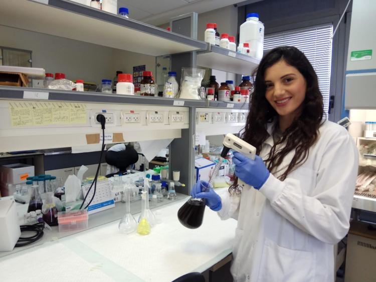
Doctoral student Chen Tzror-Azankot of Prof. Rachela Popovtzer’s is conducting research that allows to clearly distinguish between tumors and inflammation and has significant implications on the ability to diagnose – and later treat – cancer.
Last month, the prestigious ACS Nano journal – one of the leading academic publications on nano-technology – published an article titled Glucose-Functionalized Liposomes for Reducing False Positives in Cancer Diagnosis. It was written by doctoral student Chen Tzror-Azankot (31) of the nano-theranostics lab at the Faculty of Engineering in Bar Ian University. The article details her main doctoral project: developing nano-particles that help with differential diagnosis of tumors and inflammations in PET-CTs. Once the article becomes a treatment protocol, it may solve the problem of false-positive recognition of inflammation as cancer after treating the tumor. “PET scans are the main scans used today for detecting cancerous tumors, but they have one key disadvantage – inflammations can also produce a positive signal, creating a problem in distinguishing tumors from inflammations,” explains Tzror-Azankot. “It’s mostly an issue for people in recovery, who are susceptible to inflammations around the tumor area, as well as other areas of the body, as a result of the treatment; because of that we can’t always tell if the cancer is back, or if it’s just an inflammation and the patient is actually cancer-free.”
The goal of the particles is to help with this differential diagnosis. “In PET scans today we use a certain contrasting material – a radioactively marked sugar molecule. Because cancer cells consume sugar at high volumes, the tumor area would show high rates of sugar consumption which we can identify in the scan,” says Tzror-Azankot. In order to differentiate between the two, Tzror-Azankot and her team employed the characteristics of the tumor itself: the tumor, as a result of its rapid development and growth, has damaged blood vessels. This phenomenon is known as EPR (Enhanced permeability and retention). This means that the tumor is highly penetrable – it can be easily accessed through the bloodstream. “Our particles are liposomes that are also marked by sugar and injected directly into the bloodstream. But because of the size of the nano-particles, unlike the contrasting material usually used in a scan, they can exit the blood vessels only around the tumor and not inflammation. Our method therefore clearly distinguishes between tumors and inflammations in PET scans.”
Chen Tzror-Azankot, married with two children, is currently in the third year of her doctoral studies in the bioengineering track, under the supervision of Prof. Rachela Popovtzer. Before arriving at the Faculty of Engineering, first for her masters and then at the integrated doctoral track, she completed her bachelor’s degree at the Technion where she majored in biochemical engineering and then worked in the industry. “The Faculty of Engineering impressed me in terms of high professionalism and warmth, fun, and sense of family; I really enjoy studying and working here,” she says.
Tzror-Azankot’s work earned her the Ze’ev Jabotinsky scholarship for students in the direct PhD track. The scholarship is awarded by the Israel Science Foundation. “The scholarship gives me peace of mind and allows me to focus solely on my research,” she says. Her research advanced to other avenues, with the goal of “making this technology not only diagnostic but also apply it to treatment in addition to the medication that’s transmitted directly to the tumor, thereby allowing correct identification along with treatment. I’d like to take this opportunity to thank my team at the lab for their collaboration, particularly my supervisor, Prof. Rachela Popovtzer, for her constant help and support, and the head of the lab, Dr. Menachem Motiei, for helping at each and every step of the way.”
Last Updated Date : 14/04/2021